磁共振成像 (MRI) 是一种医学成像技术,它在不使用电离辐射的情况下,利用强磁场、无线电波和计算机技术来生成人体器官和组织的详细图像。人类长寿科技公司基于自有的磁共振序列和软件技术,不需要注入造影剂就能生成高清晰度的图像,有助于癌症、心血管疾病、代谢性疾病和神经退行性疾病的早期检测。
MRI与CT、PET检查的比较
电子计算机断层扫描 (CT) acquires multiple X‑ray images from different angles to reconstruct three-dimensional (3D) images. Because it uses X‑rays, the patient is exposed to ionizing radiation which increases their risk of DNA damage and subsequent cancer.
Positron Emission Tomography (PET) takes pictures based on the body’s metabolism. Many PET scans use a radioactive glucose injection that gives off energy in the form of gamma rays. Cancer cells, which grow quickly, are more likely to take up larger amounts of the glucose than normal cells, where it is then trapped and may be visualized by the PET scan.
Both PET and CT subject the individual to high levels of ionizing radiation whereas MRI uses non-ionizing radiation via a strong magnetic field and radio frequency energy to generate 3D images. It provides superior soft tissue contrast to CT and yields very detailed information about tissues and organs in our body without known negative side-effects.
Further, through the use of advanced full body MRI techniques, we are able to quantify important health parameters such as liver fat, visceral fat (fat that surrounds the internal organs), muscle mass, brain volume, ejection fraction (the proportion of blood volume pumped out of the heart with every contraction) and to detect critical health conditions such as aneurysms and cancer.
Whole-Body MRI at Human Longevity
Whole-body imaging for health assessment is performed in all patients at Human Longevity regardless of whether they have disease symptoms and is an important part of a proactive healthcare platform. This 3D imaging examination of the body anatomy and function can detect disease in the earliest stage.
1NOTE: Human Longevity Inc. uses full body and brain MRI and a limited CT for coronary artery calcium scoring (limited CT scanning area and time of exposure only to the heart).
Your comfort is of utmost importance to us. What to expect during your whole-body MRI scan at HLI
The MRI is a multisensory experience and takes approximately 60 – 90 minutes. You will be asked to lie flat with a series of coils or “cameras” placed around your head and body. Earbuds will be provided along with your choice of music, as well as a series of soothing visuals from which to choose. Clients experience noises of varying pitch and duration during the course of the exam, along with normal movement of the table. Clients may also experience a mild warming sensation and peripheral nerve stimulation, both of which are normal biological responses to full body MRI scans. Clients will be in constant contact with a technologist throughout the entire exam.
* If you are claustrophobic, we recommend bringing oral sedation. We do not advise driving after taking a sedative for at least six hours. Depending on your needs, we can assist in arranging transportation for you.
MRI Scan at Human Longevity
After your full body MRI scan, two board certified radiologists evaluate the brain and whole-body MRI and a board-certified cardiologist evaluates the cardiac MRI.
You will receive your imaging report along with your blood chemistry results one week after the exam or sooner. If there is a significant finding, your longevity physician and radiologist will work as a team to facilitate next steps including further imaging or a consultation with a specialist.
As a 100+ member, you can expect repeat annual whole-body MRI scans to identify disease at the earliest stage. If clinically indicated, more frequent imaging may be performed (for example, to follow MRI-derived liver and visceral fat percentages).
The dedicated brain MRI is comprised of high-resolution 3D anatomic images and fully automated segmentation and quantitative analysis of the 3D images using machine learning. The advantage of brain segmentation is that it provides objective clinical data to the clinical team in the form of automatically derived volumetric values and color-coded brain structures to assess and monitor biomarkers of neurodegenerative brain disorders. The movie shows about 75% of coronal slices of the color-coded brain structures from front to back, with the rest removed for de-identification.
The non-contrast angiogram examines the brain’s blood vessels and can find brain aneurysms as small as 3 millimeters in diameter. The movie shows healthy vessels including internal carotid artery (where one would take a carotid pulse) and cerebral arteries, rotating in clockwise direction to show the 360-degrees view. The middle cerebral artery, which is located in the middle of the movie extending to right and left, is the main artery that typically occludes in a large stroke.
The cardiac MRI includes images of multiple views of heart to examine function, tissue characterization and cardiac valves. The movie shows the four-chamber (right and left ventricles and atria) view of the beating heart. How much of the blood in the left ventricle gets ejected on each beat is called the ejection fraction. An ejection fraction of 60% means that 60% of the blood is ejected every time the left ventricle contracts. A cardiologist reviews and reports ejection fraction along with other quantitative cardiac biomarkers at the Human Longevity.
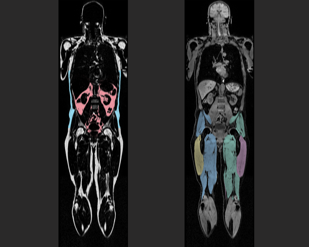
This particular whole-body MRI sequence was developed to achieve segmentation and quantification of the muscle and fat tissues. The person’s body composition profile provides quantitative imaging results to evaluate the risk for diabetes, cardiovascular disease and metabolic syndrome. Human Longevity has unique and unparalleled expertise in whole body interpretation for metabolic, vascular, neurodegenerative, cancer and cardiovascular diseases and conditions.
Photo on the left: Pink is visceral fat. Photo on the right: Different colors represent different muscles.
Fat images are segmented into visceral adipose tissue (sometimes referred to as internal belly fat) and abdominal subcutaneous adipose tissue. Visceral adipose tissue is not the unattractive superficial fat, but the inner fat that surrounds our organs. This is actually known to be more harmful than visible fat. This is body fat that is stored within the abdominal cavity and is therefore stored around a number of important internal organs such as the liver, pancreas, kidneys and intestines. Storing higher amounts of visceral fat is associated with increased risks for cardiovascular disease, stroke and type II diabetes.
Muscle images are segmented into left anterior thigh, left posterior thigh, right anterior thigh, and right posterior thigh. The posterior muscle groups include the gluteus, iliacus, adductor, and hamstring muscles among others. The anterior thigh includes quadriceps femoris, sartorius, and tensor fascia latae muscles among others.
(正文结束)
点击下载我们的全身MRI 白皮书:Full Body Magnetic Resonance Imaging from Human Longevity